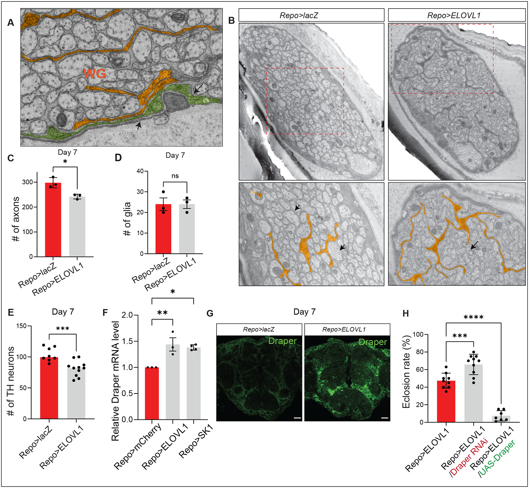
Figure 4. Elevated S1P induces phagocytosis through Draper.

(A) Image of a section of the wing nerve showing the three types of glia: Perineurial (PG), Subperineurial (SPG), and wrapping glia (WG). (B) Glial ELOVL1 expression leads to extended and expanded glial membranes. (C) The number of axons is significantly reduced in the wing margin nerves of Repo>ELOVL1 flies at Day 7 (n=3 per each genotype), however the (D) number of glia is not altered (E) The number of TH neurons is significantly reduced in CNS of Repo>ELOVL1 flies (n=6 for Repo>lacZ, n=11 for Repo>ELOVL1). (F) The relative Draper mRNA levels are significantly increased in the CNS of Repo>ELOVL1 and Repo>SK1 flies (n=3 per each genotype). (G) Draper protein levels are increased in Repo>ELOVL1 fly heads. (H) Eclosion rates of Repo>ELOVL1 flies are modulated by Draper expression. Quantification of the percentage of expected animals per cross (n>7). Statistical analyses are one-way ANOVA followed by a Tukey post hoc test. Results are mean ± s.e.m. (****p < 0.0001, ***p < 0.001, **p < 0.01; n.s., not significant).
