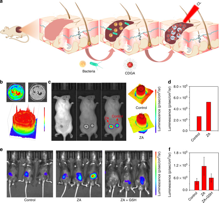
Fig. 4. In vitro and in vivo bioimaging of bacteria-associated inflammation.
a Schematic illustration of the CL sensing in inflammation model. b The CL emission of the CDGA recorded by the IVIS system. c The CL images of mice treated with and without the ZA in superficial wound. d The quantification of corresponding CL intensity from the images of superficial wound. e The CL images of mice intraperitoneally treated with ZA, ZA plus GSH and saline, following by an intraperitoneal injection of CDGA at t = 4 h. f The quantification of corresponding CL intensity from the in vivo CL images

