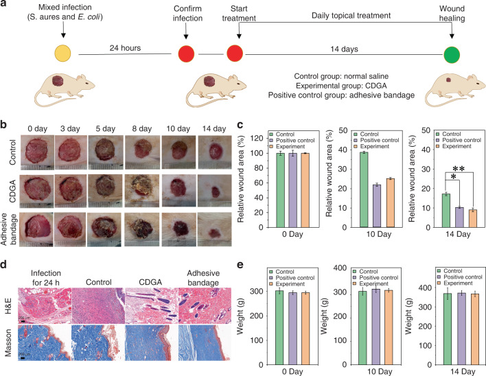
Fig. 6. In vivo antibacterial capability of the CDGA.
a The experimental process of mice trauma model with the mixed infection of S. aureus and E. coli. b The photographs of the infected wounds in negative control group (Control, normal saline), experimental group (CDGA), and positive control group (Adhesive bandage). c The relative wound area at 0, 10 and 14 days for different groups. (*p < 0.05, **p < 0.01). d The images of H&E staining and Masson’s staining, including infection for 24 h and treatment for 14 days. Scale bar = 200 μm. e The body weight variations at 0, 10 and 14 days for different groups

