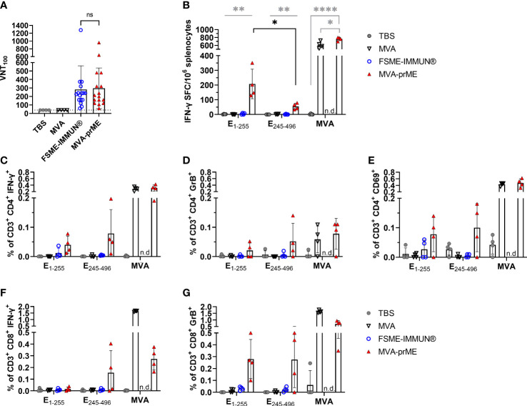
Figure 2.
Humoral and cellular immune response in vaccinated mice. (A) Virus-neutralizing titer (VNT100) against TBEV Neudoerfl of murine sera samples obtained 56 days after prime immunization (samples of immunogenicity and protective efficacy study, n = 16). Samples with ≤40 VNT100 (dotted line, lowest serum dilution) are considered negative. The FSME-IMMUN®-vaccinated mouse that displayed signs of disease is highlighted with a diamond symbol. n.s., not significant (p>0.05). (B) Displayed are IFN-γ spot-forming cells (SFC) per one million splenocytes after background subtraction. Only significance between MVA-prME to other treatment groups is indicated (gray) and for E1-255 versus E245-496 of MVA-prME-vaccinated mice (black) (*p≤0.05, **p≤0.01, ****p≤0.0001). (C–G) Frequency of CD3+ subpopulations gated on CD4+IFN-γ+ (C), CD4+Granzyme B+ (D), CD4+CD69+ (E), CD8+IFN-γ+ (F), or CD8+Granzyme B+ (G) after background substraction. n.d., not determined. For all graphs, bars show the mean with standard deviation. Mice were either immunized with TBS (gray circle), MVA (non-filled triangle), FSME-IMMUN® (non-filled blue circle), or MVA-prME (red triangle).

