Figure 5.
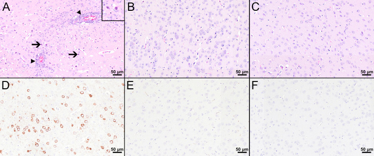
Histological and immunohistochemical analyses of the cerebral cortex at 8 dpi. (A–C) H&E-stained sections of the cerebral cortex of TBEV-infected mice which were either treated with PBS (A) or vaccinated with FSME-IMMUN® (B) or MVA-prME (C). (A) The cerebral cortex of the PBS-treated mouse displays cellular necrosis with karyorrhectic, karyolytic, and pyknotic cells (insert) and shrunken, hypereosinophilic, triangular-shaped necrotic neurons (arrows and insert) as well as inflammatory cell infiltrates in destructed vascular walls (necrotizing vasculitis; arrowheads) and the perivascular space (arrowheads). Microgliosis and hypertrophy of microglia/macrophages are present. (B, C) In FSME-IMMUN®- (B) or MVA-prME-vaccinated (C) mice, no significant microscopic lesions within the cerebral cortex parenchyma are visible. (D–F) IHC for the TBEV E antigen of the cerebral cortex of TBEV-infected mice which were either treated with PBS (D) or vaccinated with FSME-IMMUN® (E) or MVA-prME (F). (D) Immunohistochemically, a cytoplasmic TBEV immunoreactivity is present in multiple cells representing neurons of the cerebral cortex from a PBS-treated mouse. (E, F) The cerebral cortex of FSME-IMMUN®- (E) and MVA-prME-vaccinated (F) mice lack immunoreactivity. Scale bars: 50 µM.

