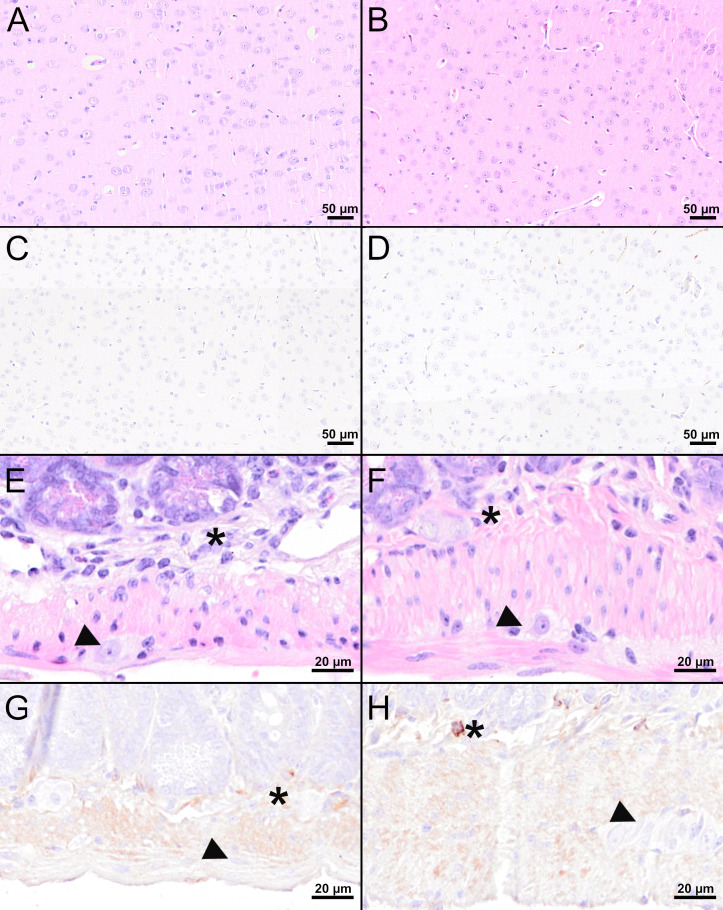
Figure 7.
Histological and immunohistochemical analyses of the cerebral cortex and jejunum at 16 dpi. (A, B, E, F) H&E-stained sections of the cerebral cortex (A, B) and jejunum (E, F) of TBEV-infected mice which were either vaccinated with FSME-IMMUN® (A, E) or MVA-prME (B, F). (C, D, G, H) IHC for TBEV E antigen of the cerebral cortex (C, D) and jejunum (G, H) of TBEV-infected mice either vaccinated with FSME-IMMUN® (C, G) or MVA-prME (D, H). (A, B) No significant microscopic lesions within the cerebral cortex parenchyma are visible. (C, D) TBEV immunoreactivity was absent within the parenchyma of the cerebral cortex. (E, F) The jejunum shows mild hypercellularity of the submucosal plexus (asterisk), while the myenteric plexus (arrowhead) reveals no significant findings. (G) No specific TBEV immunoreactivity is detectable in the submucosal (asterisk) or the myenteric plexus (arrowhead). (H) A single cell in the submucosal plexus (asterisk) is immunolabeled, while no immunoreactivity is present in the myenteric plexus (arrowhead). Scale bars: (A–D) 50 µM, (E–H): 20 µM.

