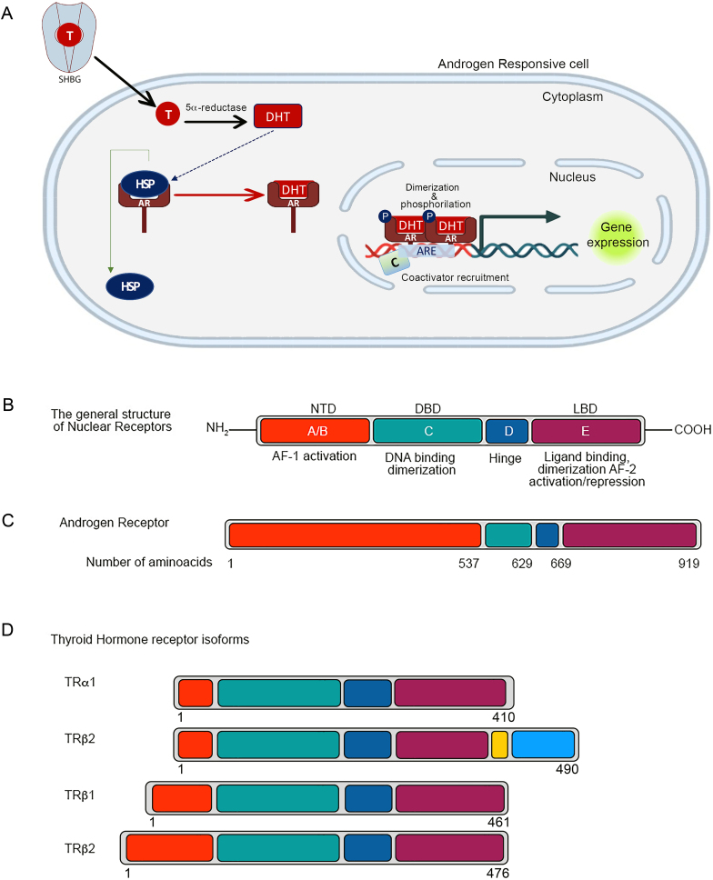
Figure 1.
Mechanism of action of androgen receptor and structure of androgen receptor (AR) and thyroid receptor. (A) AR action. After dissociation from the carrier (SHBG) and entering the cell, free testosterone is metabolized into 5α-dihydrotestosterone (DHT) by the enzymatic action of 5α-reductase. DHT binds to the AR and dissociates it from heat-shock protein (HSP). The androgen/AR complex translocates to the nucleus, dimerizes, and binds to the promoter of the target genes (ARE). Then, the complex recruits coactivators and other transcription factors (coregulator proteins) to modulate gene transcription. (B) Nuclear receptors comprise four sections, including the A/B region which consists of the N-terminal domain (NTD) and the first transactivation domain (AF-1); the C region or the DNA-binding domain (DBD); the D domain known as the hinge region, which promotes DBD binding to DNA; and the E region including C-terminal ligand-binding domain (LBD) and the second transactivation domain (AF-2). (C) The structure of the AR containing the regions enumerated for nuclear receptors as a whole. (D) The structure of four thyroid hormone isoforms. TRβ1 and TRβ2, the most known TR isoforms, differ in the NTD, but TRα1 and TRα2 vary in the LBD. TRα2 with longer LBD is unable to bind to the thyroid hormones. The digits indicate the number of beginning and ending human amino acids in each section. AF-1 activation domain is indicated in red, the DBD is indicated in green; the hinge domain is indicated in dark blue; the LBD is indicated in violet and TRα2 carboxy termini are indicated in yellow and light blue. AR, androgen receptor; DBD, DNA-binding domain, DHT, dihydrotestosterone; HSP, heat-shock protein; LBD, ligand-binding domain; NTD: N-Terminal Domain; SHBG, sex hormone binding globulin.

 This work is licensed under a