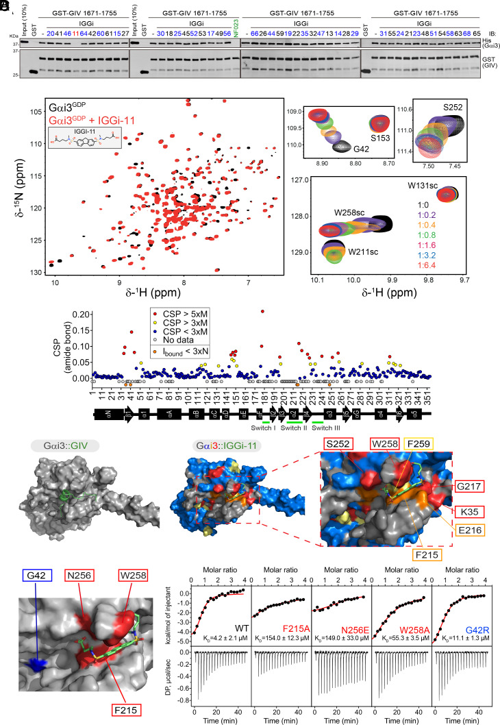
Fig. 2.
IGGi-11 binding to the GIV-interacting region of Gαi. (A) IGGi-11 disrupts GIV–Gαi binding in pull-down assays. His-Gαi3 was incubated with glutathione agarose-bound GST–GIV (aa 1671-1755) in the presence of the indicated compounds or the positive control NF023 at a concentration of 100 μM. After incubation and washes, bead-bound proteins were separated by SDS-PAGE and immunoblotted (IB) as indicated. Representative of 3 independent experiments. (B) Overlay of 1H–15N TROSY spectra of 2H,13C,15N–Gαi3–GDP in the absence or presence of IGGi-11. Selected regions from the overlaid spectra depicting representative perturbations in Gαi3 signals induced by increasing amounts of IGGI-11 are shown on the right. The scatter plot (bottom) corresponds to the quantification of IGGi-11-induced chemical shift perturbations (CSPs). Red, CSP > 5 times the median (M); yellow, CSP > 3xM; blue, CSP < 3xM; gray, no data. Reductions in signal intensity (Ibound) below three times the noise (N) are indicated in orange. (C) Comparison of models of IGGi-11 docked onto Gαi3 (Middle and Right, color coded according to NMR perturbations quantified in A) and GIV-bound Gαi3 (Left). (D) Quantification of IGGi-11 binding affinity (KD) for Gαi3 wild type (WT) or the indicated mutants using isothermal titration calorimetry (ITC). Data are representative of at least two independent experiments.

