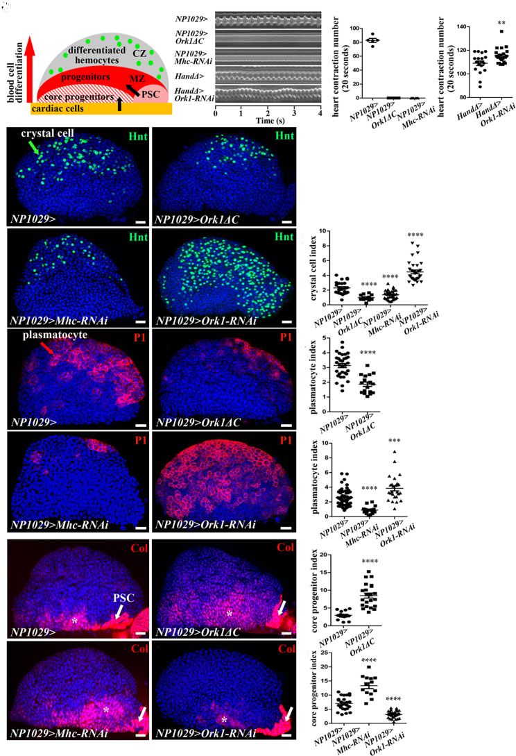
Fig. 1.
Heartbeat controls lymph gland homeostasis. (A) Schematic representation of a third-instar lymph gland anterior lobe. It is composed of progenitors (red) and core progenitors (hatched red) in a medullary zone (MZ), and a cortical zone, which contains differentiated hemocytes (CZ, green dots). The two niches, the PSC (pink) and the cardiac cells (orange), regulate different subsets of MZ progenitors (black arrows). (B) Kymograph of heartbeat in the control (NP1029> or HandΔ>) and when Ork1ΔC or Mhc-RNAi, or Ork1-RNAi are expressed with cardiac cell drivers. (C and D) Number of heart contractions per 20s. (E–H) Crystal cell differentiation (Hnt, green) when the heart is blocked (F and G) or accelerated (H). (I) Crystal cell index. (J, K, M, and N) Plasmatocyte differentiation (P1, red) when the heart is blocked (K and M) or accelerated (N). (L and O) Plasmatocyte index. (P, Q, S, and T) Col (red) labels core progenitors (*) and the PSC (arrow). (R and U) Coreprogenitor index. For all quantifications and figures, statistical analysis t test (Mann–Whitney nonparametric test) performed using GraphPad Prism 5 software. Error bars represent SEM and *P < 0.1; **P < 0.01; ***P < 0.001; ****P < 0.0001. ns (not significant). Nuclei are labeled with Topro (blue). (Scale bars, 20 µm.)

