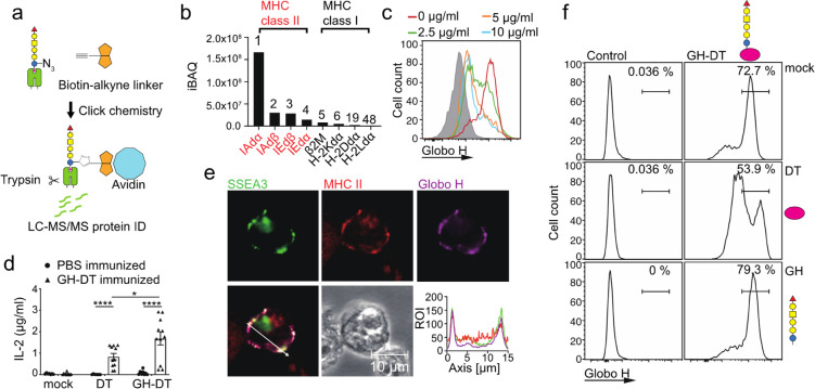
Figure 4.
GH and SSEA3 glycans were presented through MHC class II on BMDCs treated with GHN3-DT. (a) Flow chart for isolation and identification of proteins interacting with GHN3 glycan and intermediates during antigen presentation in GHN3-DT-treated BMDCs. The proteins interacting with GHN3 and its intermediates in complex with MHC class II were isolated for LC–MS/MS analysis. The azido groups of the complex were first biotinylated with click reaction followed by treatment with avidin beads to pull down the interacting proteins in the complex. (b) Among the 1231 identified proteins, 194 proteins were membrane proteins and MHC class II was the most dominant hit. All of MHC class I subunits were also identified. (c) Addition of anti-MHC class II antibody reduced the presentation of GH glycan on BMDCs 48 h after GH-DT treatment. (d) Compared to DT, GH-DT induced more IL-2 production from CD4+ T cells isolated from GH-DT-immunized mice. (e) GH and SSEA3 glycans were colocalized with MHC class II on the surface of GH-DT-treated BMDCs. Cells were harvested and stained 24 h after GH-DT treatment. Shown is the distribution of SSEA3 glycan (green), MHC class II (red), and GH glycan (purple) indicated by fluorescence intensities across the section with a white line. (f) Effect of DT (1 mg/mL) or GH glycan (1 mg/mL) on antigen presentation of GH-DT-treated BMDCs. Results in (d) are mean ± SEM (n = 11). * p < 0.05, **** p < 0.0001.

