Figure 4.
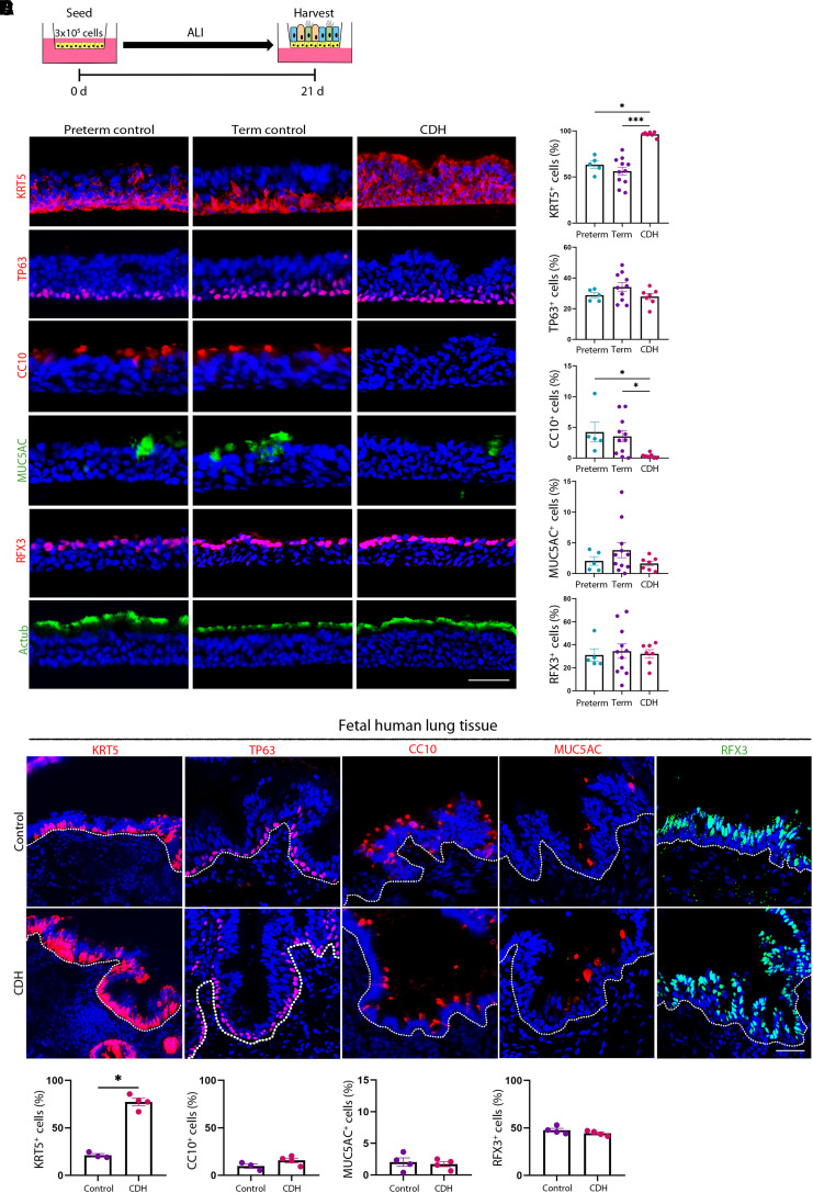
Congenital diaphragmatic hernia (CDH) basal stem cells (BSCs) have similar epithelial differentiation defects in vitro and in vivo. (A) Schematic of air–liquid interface (ALI) culture of preterm, term, and CDH BSCs for 21 days. (B) Representative fluorescence images of staining for markers of BSCs (KRT5 and TP63), club cells (CC10), goblet cells (MUC5AC), and ciliated cells (RFX3 and Actub) using cross-sections of fixed ALI cultures. Scale bar, 25 μM. (C) Quantification of the relative abundance of different epithelial cell types on the basis of the staining of ALI culture in triplicates. More than 6,000 nuclei in five 20× images were counted for each marker for each ALI culture. Each dot represents one BSC line. (D) Representative fluorescence images of immunostaining for KRT5, TP63, CC10, MUC5AC, and RFX3 and in term, control (n = 3), and CDH (n = 4) human fetal lung sections. Scale bar, 50 μm. More than 2,000 nuclei in five images were counted per sample. (E) Quantification of the relative abundance of epithelial cells expressing individual markers on the basis of the staining of human fetal lung sections. Bar graphs show mean ± SEM. Statistical analyses were performed using the Kruskal-Wallis test for comparisons of multiple experimental groups and the Mann-Whitney U test for comparisons between two groups. Significance indicated as *P < 0.05 and ***P < 0.001.

