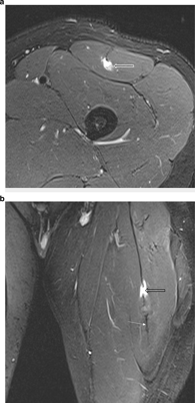
Figure 10.
A 27-year-old professional male footballer with a BAMIC Grade 2c injury of the left rectus femoris which appears to involve the capsular head in isolation. (a) Axial T2FS. Note the focal oedema around the deep portion of the intramuscular myotendon with absence of the normal tendon fibres. Also note the relative sparing of the remaining indirect head tendon which has a normal morphology. As suggested by Tubbs et al, this is the region of the capsular head fibres suggesting isolated capsular head tear. (b) Coronal STIR sequence of same athlete as Figure 10a demonstrating complete disruption of tendon fibres within the deep-most fibres of the intramuscular tendon (black open arrow). Note scarring of the more distal tendon fibres in keeping with recurrent injury (arrow). STIR, short tau inversion-recovery

