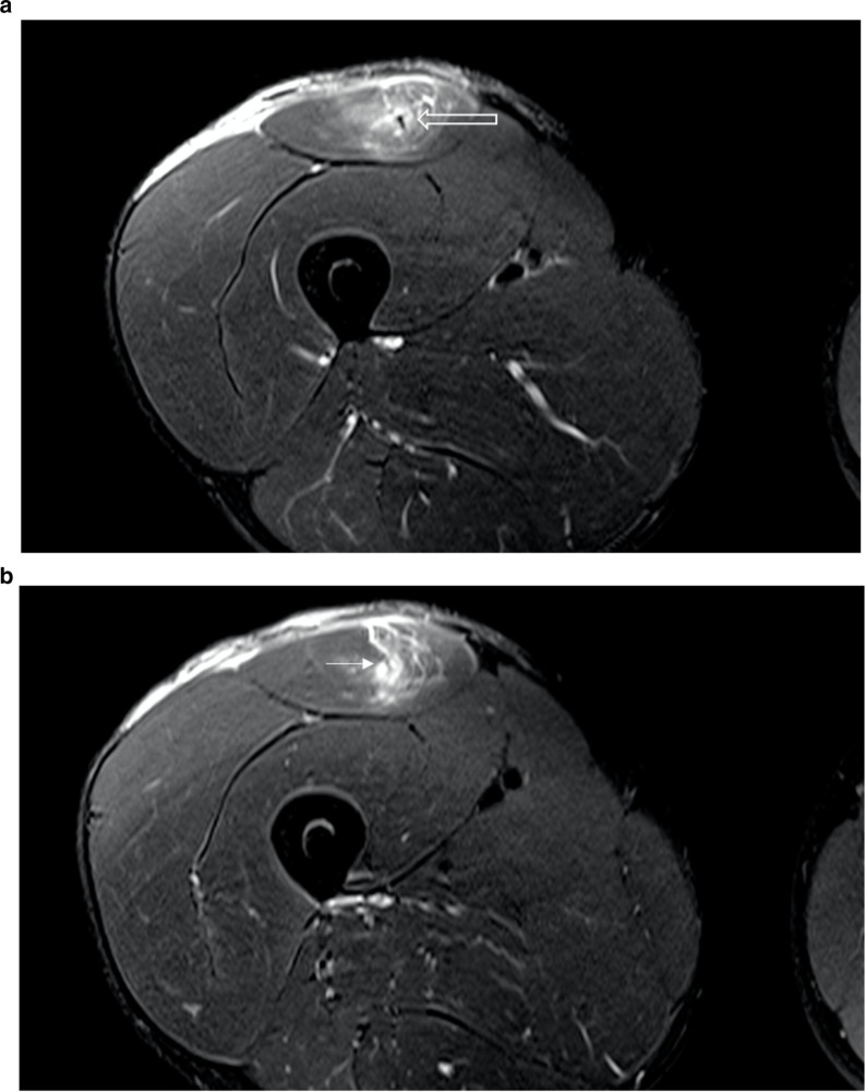
Figure 7.
A 22-year-old professional male footballer with a BAMIC Grade 3c injury of the proximal right rectus femoris. (a) Axial T2FS. Note the full-thickness tear of the direct and indirect heads. Again, the presence of the likely capsular head component of the deep myotendon is noted (open arrow). (b) Axial T2FS. Distal to Figure 7a the capsular head now demonstrates full-thickness rupture with fluid replacing the entirety of the deep myotendinous junction of the proximal rectus femoris (arrow). (c) Axial T2FS. Distal to Figure 7b the intramuscular tendon re-emerges with the linear-appearing indirect head (arrow) and the slightly thickened rounded capsular head (open arrow) lying within the deeper aspect of the fibres (tadpole sign).

