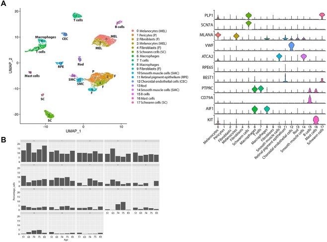
Figure 3.
Single-cell RNA-Seq of adult human RPE–choroid tissue. (A) Integrated UMAP (left) revealing the presence of 18 cell clusters in the adult RPE–choroid tissue and violin plots (right) showing the expression of key cell type specific markers. (B) Cell type representation in RPE–choroid tissue across five different donors of different ages. The age is shown on the x axis and cluster number on top of each panel.

