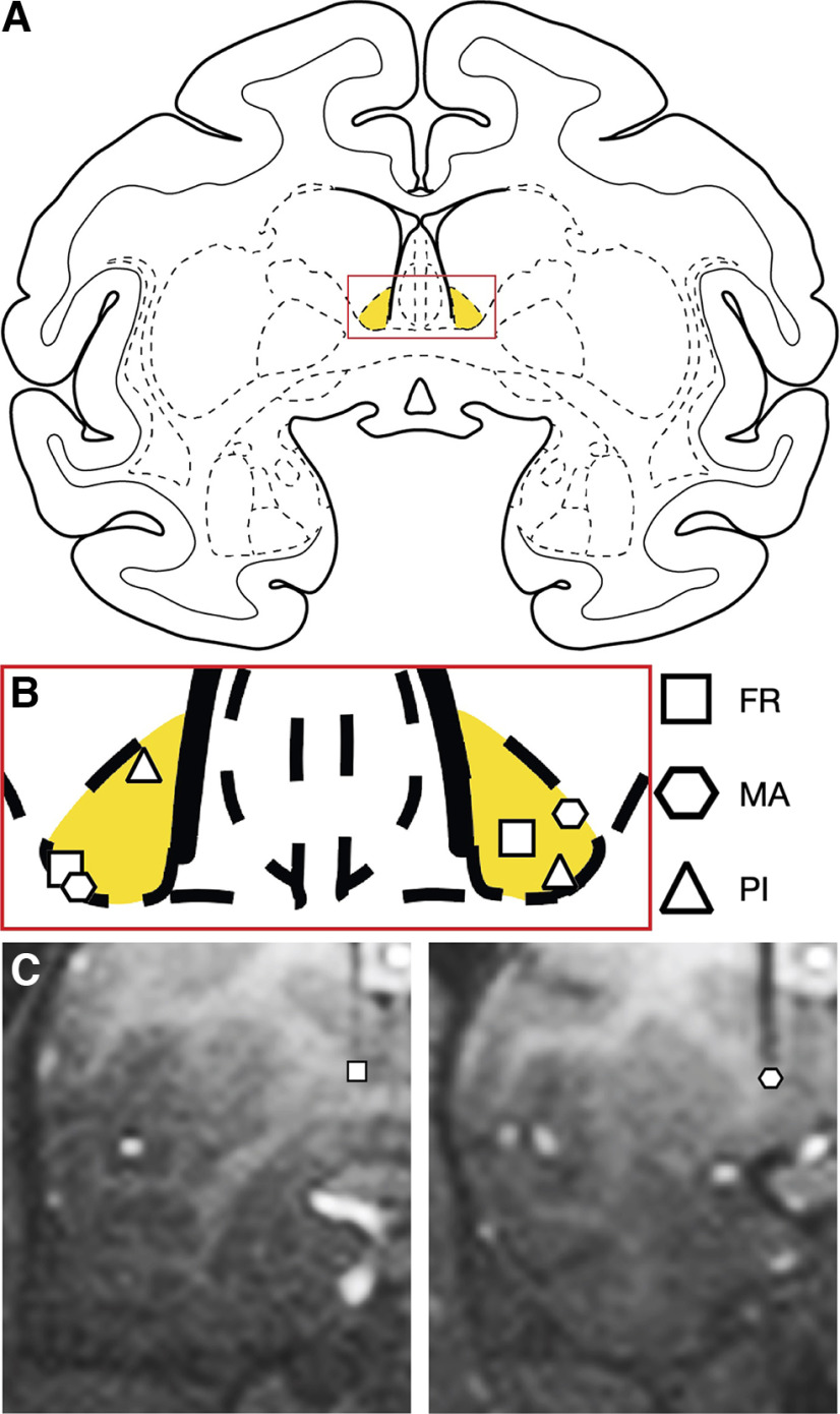
Figure 1.
A, A coronal atlas plane showing the area of intended drug infusion. Yellow area represents the anterior BNST. B, Placement of individual infusions in the 3 subjects based on postoperative MRI. C, MRI images from 2 subjects showing the placement of a tungsten microelectrode at the dorsal border of the BNST.

