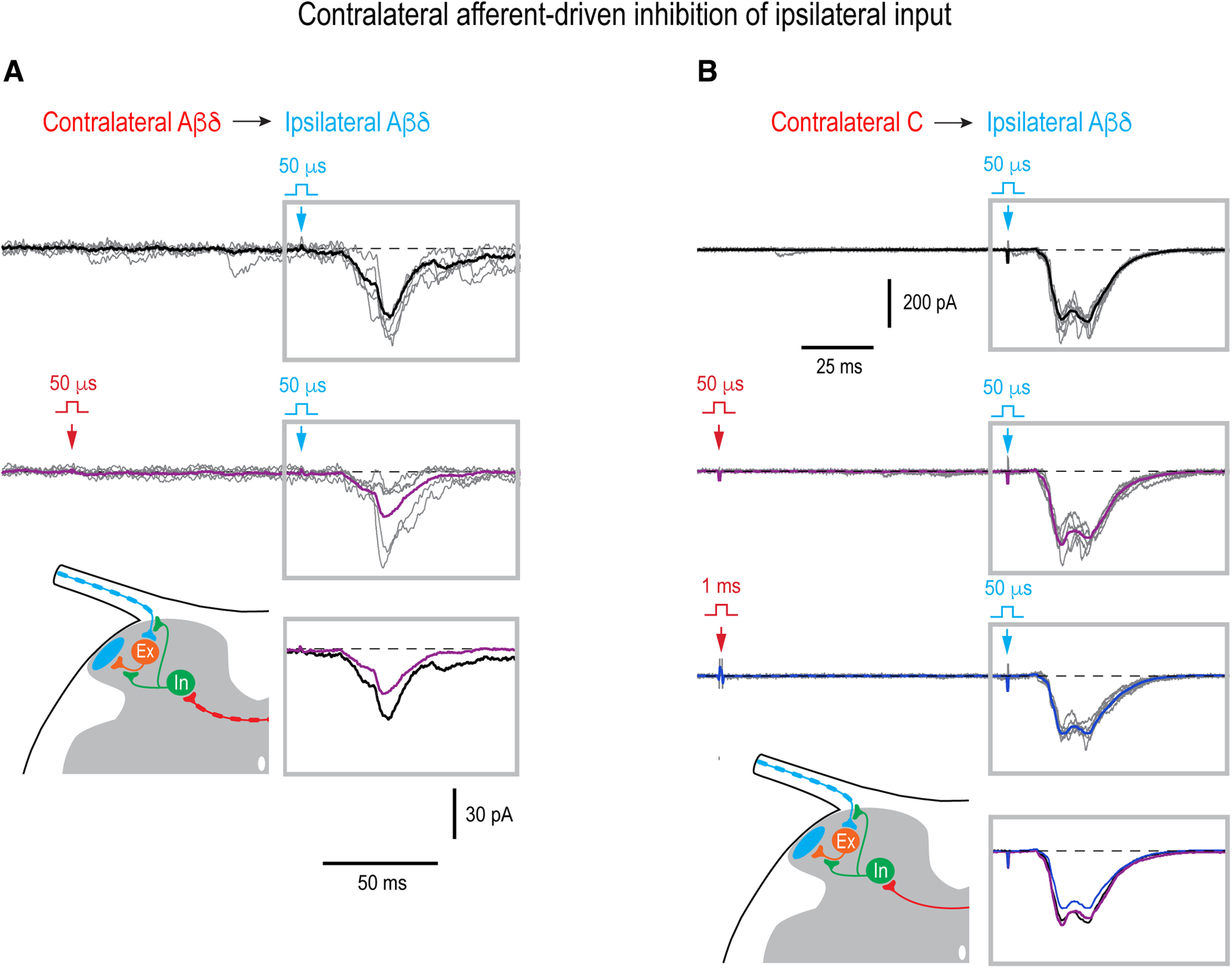
Figure 7.

Contralateral control of the ipsilateral overall input. A, Contralateral Aβδ-range conditioning attenuated the ipsilateral Aδ-fiber input in a PN (integral reduced by 55%, p = 0.03, unpaired t test). Holding potential, −70 mV. B, Contralateral Aβδ-conditioning did not change the ipsilateral Aδ-fiber input (p = 0.3, unpaired t test) that was however significantly attenuated by the contralateral Aβδ/C-conditioning (integral reduced by 21%, p < 0.001, unpaired t test). Thus, the effect was considered to be driven by the contralateral C-afferents. Holding potential, −80 mV. In A, B, 50-µs stimuli were applied to activate Aβδ-afferents and 1-ms stimuli to activate all Aβδ/C-afferents. The time interval between the contralateral conditioning stimulus (red arrow) and ipsilateral test stimulus (blue arrow) was 100 ms. For each type of response, five traces are shown with the average of 7–21 traces. Averaged responses to the test stimuli are shown superimposed below. Schematics show contralateral Aβδ-afferent-driven (A) and C-afferent-driven (B) presynaptic inhibition of the ipsilateral afferent supplying an intercalated excitatory neuron or/and of the axon terminal of the intercalated neuron. The presynaptic, rather than postsynaptic, mechanism of the contralateral inhibition of the ipsilateral polysynaptic input was assumed, since effect was observed 100 ms after conditioning stimulation when cDRP, and therefore PAD, reached its maximum (Fig. 6A), but most evoked IPSCs already terminated (Luz et al., 2019; Fernandes et al., 2022a). Locations of inhibitory (In) and excitatory (Ex) interneurons are not known.
