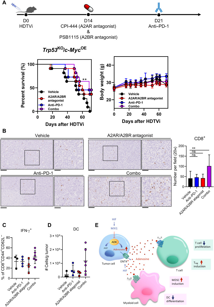
Fig. 7. Adenosine receptors blockade synergizes with anti–PD-1 treatment.
(A) In vivo HCC tumors were induced via HDTVi of plasmids carrying Trp53KO/c-MycOE in C57BL/6N mice. (Left) Survival plot and (right) body weight of HCC-bearing mice treated with vehicle, adenosine receptor antagonists, anti–PD-1 monoclonal antibodies, or the combination of both. (B) Representative pictures (left) and quantification (right) of CD8+ T cells in tumor by immunohistochemistry staining. (C) Percentage of effector T cells (CD8+CD44+CD62L−) expressing IFN-γ. (D) Quantification of DCs in tumor by flow cytometry. (E) Schematic summary of the role of HIF in promoting intracellular adenosine efflux and mediating immunosuppressive TME. Error bars indicate means ± SD. **P < 0.01 versus vehicle. (A) Kaplan-Meier followed by log-rank test. (B) Student’s t test. Original, 20× magnification (scale bars, 100 μm); Inset, 40× magnification (scale bars, 50 μm).

