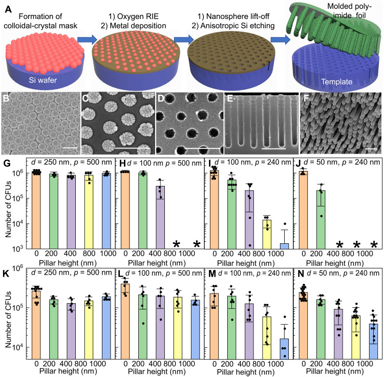
Fig. 2. Biomimetic mechano-bactericidal nanopillar arrays on the outer surface of the dual-functional smart-coating foil protecting orthopedic implants against bacterial infections.
(A) Schematics depicting the process to prepare polyimide foils featuring nanopillar arrays with tunable structures. (B to F) Scanning electron microscopy (SEM) images showing the critical steps: Top-view SEMs of the colloidal-crystal mask before (B) and after (C) oxygen RIE, and the metal hard mask formed after lift-off (D); cross-sectional SEM micrograph of the template after deep-silicon RIE (E); and tilted-view SEM showing the representative nanopillar arrays on the prepared polyimide foil (F). Scale bars, 400 nm. (G to J) Counts of viable E. coli (MG1655) after being incubated with 1-cm by 1-cm nanopillar arrays with varying nanopillar diameter (d), pitch (p), and height (h) and planar controls for 90 min. Asterisk indicates the number of colony-forming units (CFUs) was less than 1000. (K to N) Counts of viable MRSA (USA300) after being incubated with 1-cm2 nanopillar arrays and planar controls. p = 500 nm and d = 250 nm for (G) and (K); p = 500 nm and d = 100 nm for (H) and (L); p = 240 nm and d = 100 nm for (I) and (M); and p = 240 nm and d = 50 nm for (J) and (N). Zero height represents the planar foils as internal controls included in each experiment. N ≥ 4 in all experiments.

