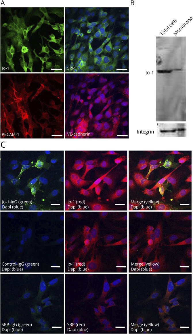
Figure 5. Jo-1 Antibodies Bound to Jo-1 in TSM15 Cells.
(A) Immunohistochemical staining of Jo-1 and SRP in TSM15 cells. Platelet endothelial cell adhesion molecule 1 and vascular endothelia-cadherin are stained in red; nuclei are counterstained with DAPI in blue. (B) In TSM15 cells, Western blotting demonstrated the band of Jo-1 in total cell lysis and cell membrane fraction. (C) Immunofluorescence labeling of TSM15 cells with IgG from patients with Jo-1 antibody–positive myositis (500 μg/mL) (green) or SRP antibody–positive myositis (500 μg/mL) (green) and commercial anti–Jo-1 antibodies (red) or anti-SRP antibodies (red) shows the colocalization of anti–Jo-1 antibodies and Jo-1 (merged in yellow). Scale bar, 50 μm. SRP = signal recognition particle.

