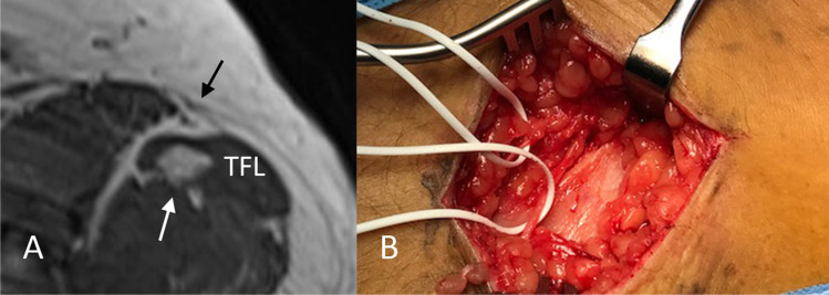
Fig. 3.
Case of lipoma inside the tensor fascia latae muscle. A: Transverse T1-weighted MR image showing a lesion of 1cm in diameter in the tensor fascia latae muscle (TFL) with high signal intensity comparable to that of subcutaneous fat (white arrow). The black arrow points at the branches of the LFCN. B: Intra-operative image with white bands placed around the separate branches of the LFCN after opening of the fascia lata

