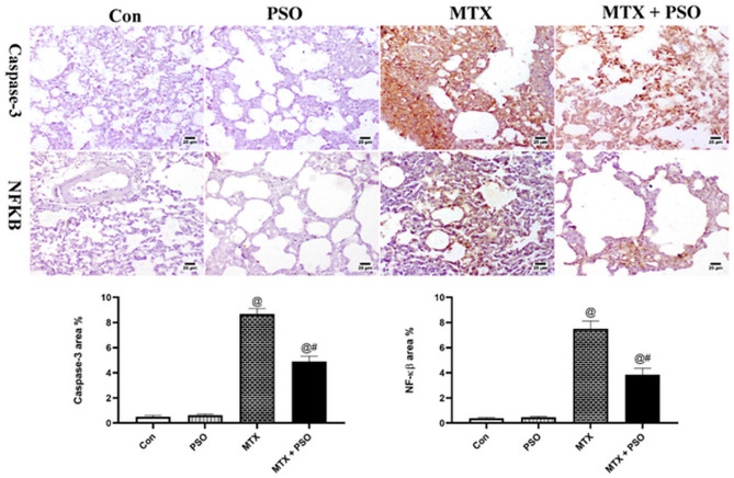
Figure 5.
Immunohistochemical staining of caspase-3 and NF-kβ in different groups. Control and PSO groups showed limited to negative expression in both markers. MTX showed strong positive expression in the inflamed lung sections concerning caspase-3 and NF-kβ. Remarkable decrease in immune staining is detected in MTX + PSO group of both caspase-3 and NF-kβ. Charts represent area % expression of immune markers in different groups. Values are expressed as means ± SE. @ significant from Con, # significant from MTX. Significant difference is considered at p < 0.05.

