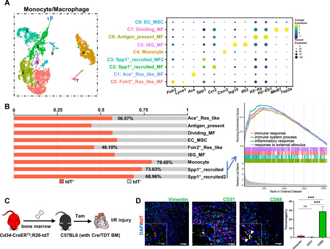
Fig. 5.
Contribution of Cd34-lineage monocytes/macrophages in I/R injury. A UMAP plot showing 9 color-coded subclusters from the monocytes/macrophages population in I/R injury, n = 4579 cells. Dot plot shown on the right indicating the expression of selected marker genes across all subclusters, with putative cell identities annotated in the middle. B Proportion of tdT+ cells in each monocyte/macrophage subcluster. Gene set enrichment analysis (GSEA) shown on the right indicated significant pathways enriched in recruited subclusters (monocytes, Spp1+ macrophages). C Sketch of the experimental design for generating chimeric C57BL6 mice with bone marrow cells transplanted from Cre/TDT mice. Chimeric mice were subjected for I/R injury and heart samples were harvested for further investigation. D Representative images of chimeric C57BL6 murine ventricles staining tdT with vimentin, CD31 and CD68 in border zones, with magnification of the boxed region, arrows indicated co-staining cells. Scale bars, 100 μm, and 20 μm in magnification. Quantification of the percentages of vimentin+/CD31+/CD68+ cells in BM-derived tdTomato+ cells by immunofluorescence was shown on the right panel. N = 4 mice, ***P < 0.001, by one-way ANNOVA test. BM bone marrow, ISG interferon stimulated, tdT tdTomato

