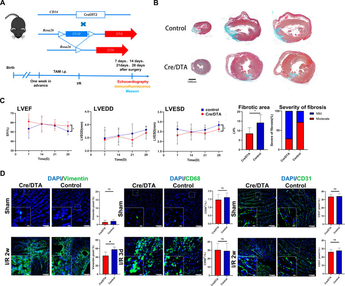
Fig. 7.
Pathological impact of Cd34-lineage cells on ischemic heart. A Experimental scheme whereby Cd34-CreERT2;R26-DTA (Cre/DTA) or control mice were given tamoxifen for 1 weeks before I/R injury. Hearts were harvested at 7, 14, 21, and 28 days after surgery. B Masson staining showing cross sections of different horizontal planes (apex, left ventricles) of hearts from Cre/DTA or control mice at 28 days post-I/R, indicating different degrees of fibrosis. Scale bar, 1 mm. C Echocardiographic measurements of left ventricle ejection fraction (LVEF), left ventricle end-diastolic diameter (LVEDD) and left ventricle end-systolic diameter (LVESD) in Cre/DTA and control mice at the indicated time after I/R injury, and percentage of fibrotic area (area of scar, normalized to the area of the ventricle) along with severity of fibrosis (fraction of mice demonstrating mild or moderate scarring) after 28 days post-I/R in two mouse groups. N = 7 mice per group. D Representative images of Cre/DTA or control ventricles staining vimentin, CD68 or CD31 showing cellular changes of mesenchymal cells, macrophages or endothelial cells after CD34+ cell ablation, with magnification of the boxed region. N = 5 mice per group. Scale bars, 100 μm, and 20 μm in magnification. Data were shown as mean ± SEM, *P < 0.05, by one-way ANNOVA test or Student’s t test

