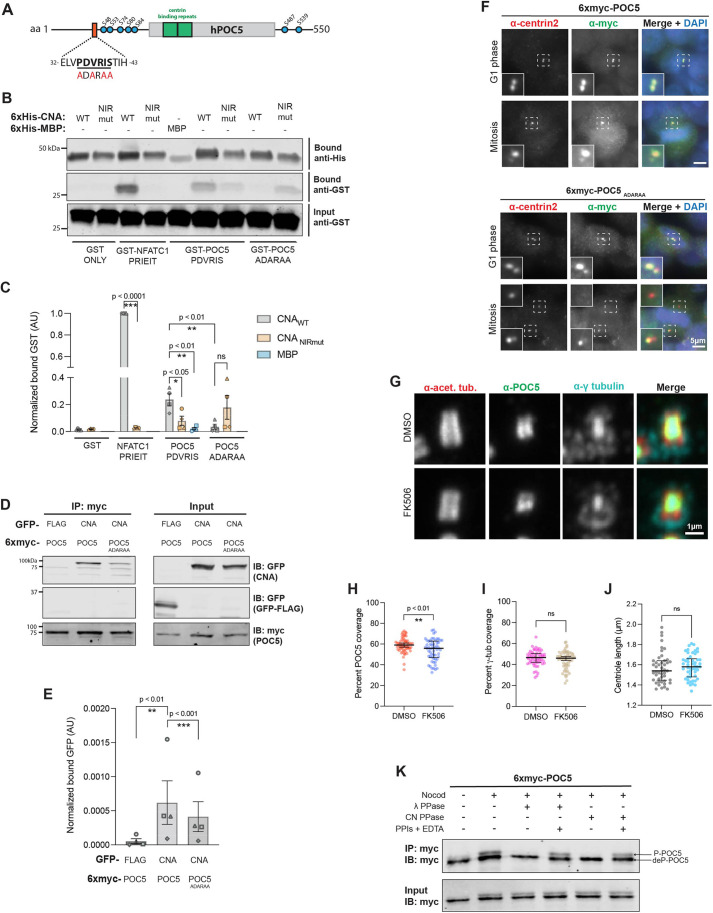
Fig. 3.
Calcineurin interacts with POC5 in a PxIxIT-dependent manner. (A) Schematic of human POC5 (hPOC5; UniProt ID Q8NA72-3). Orange, PxIxIT motif with WT (PDVRIS) and mutant (ADARAA) sequences shown beneath; green, centrin-binding repeats; blue circles, phosphorylated residues (from PhosphoSitePlus database; Hornbeck et al., 2015). (B) POC5 contains a CN-binding PxIxIT motif. Co-purification of GST-tagged PxIxIT peptides from NFATC1 and POC5, as indicated, with 6×His–MBP, 6×His–CNAαWT:CNB or 6×His–CNAαNIRmut:CNB. (C) Quantification of the experiment shown in B (bound GST signal/bound His signal, normalized to input GST signal; AU, arbitrary units). Data are mean±s.e.m. (n=4 independent experiments). ns, not significant; *P<0.05; **P<0.01; ***P<0.001 (unpaired, two-tailed Student's t-test). (D) CN association with POC5 is PxIxIT-dependent in vivo. GFP–FLAG or GFP–CNAαWT co-immunoprecipitated with 6×myc–POC5WT or 6×myc–POC5ADARAA expressed in HeLa cells. IB, immunoblot; IP, immunoprecipitation. (E) Quantification of the experiment shown in D (bound GFP signal/bound myc signal, normalized to input GFP signal; AU, arbitrary units). Data are mean±s.e.m. (n=4 independent experiments). **P<0.01; ***P<0.001 (ratio-paired, two-tailed t-test). (F) Transiently expressed 6×myc–POC5WT (top) or 6×myc–POC5ADARAA (bottom) is recruited to centrosomes in HeLa cells. Single z-plane images of cells stained for centrin 2 and myc-tagged POC5 in the indicated cell cycle stages are shown (representative of 100 cells). Dashed boxes indicate regions magnified in inset images. (G) U-ExM of centrosomes in hTERT-RPE1 cells treated with DMSO or 2.5 μM FK506 for 48 h. Maximum-intensity projections of confocal z-stacks showing staining of acetylated tubulin (acet. tub.), POC5 and γ-tubulin. (H) CN inhibition decreases POC5 luminal distribution. Percentage of centriole covered by POC5 is plotted. (I) CN inhibition does not alter γ-tubulin (γ-tub) luminal distribution. Percentage of centriole covered by γ-tubulin is plotted. In H and I, data are pooled from two independent experiments, with individual data points plotted and the median±interqurtile range (IQR) indicated. DMSO, n=60 centrioles; FK506, n=58 centrioles. ns, not significant; **P<0.01 (unpaired, two-tailed Student's t-test). (J) CN inhibition does not alter centriole length. Median±IQR centriole length is shown. Data pooled from two independent experiments. DMSO, n=46 centrioles; FK506, n=51 centrioles. ns, not significant (unpaired, two-tailed Student's t-test). (K) CN dephosphorylates mitotic POC5 in vitro. Immunoblots of in vitro dephosphorylation of 6×myc–POC5 by λ phosphatase or CN. Nocod, nocodazole synchronization; λ PPase, λ phosphatase; CN PPase, purified constitutively active truncated 6×His–CNAαWT:CNB; PPIs, phosphatase inhibitors; P-POC5, phosphorylated POC5; deP-POC5, dephosphorylated POC5. Blots shown are representative of three experiments.

