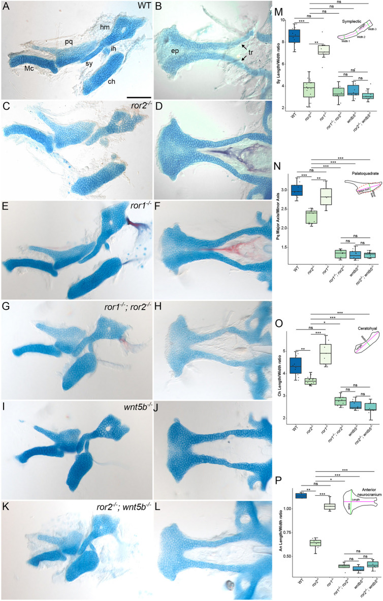
Fig. 3.
Cartilage phenotypes in ror1 and ror2 mutants. (A-L) Alcian Blue-Alizarin Red staining of craniofacial cartilages at 5 dpf. Lateral views of the upper and lower jaw cartilages (A,C,E,G,I,K) and cartilages of the anterior neurocranium (B,D,F,H,J,L). Representative images of cartilages from wild-type (WT; A,B), ror2−/− (C,D), ror1−/− (E,F), ror1−/−; ror2−/− (G,H), wnt5b−/− (I,J) and ror2−/−; wnt5b−/− (K,L) animals. Arrows point to trabecular cartilages. Anterior is to the left for all panels. (M-P) Quantifications of measured ratios: sy length-to-width (M); pq major axis-to-minor axis (N); ch length-to-width (O); An length-to-width (P). Diagrams of the sy cartilage, pq cartilage, ch cartilage and An indicating measured features are inset in panels M-P. n=10 WT, 9 ror1−/−, 18 ror2−/−, 10 ror1−/−; ror2−/−, 10 wnt5b−/− and 12 ror2−/−; wnt5b−/− sy cartilages for M. n=9 WT, 9 ror1−/−, 18 ror2−/−, 10 ror1−/−; ror2−/−, 10 wnt5b−/− and 12 ror2−/−; wnt5b−/− pq cartilages for N. n=10 WT, 9 ror1−/−, 18 ror2−/−, 10 ror1−/−; ror2−/−, 10 wnt5b−/− and 12 ror2−/−; wnt5b−/− ch cartilages for O. n=4 WT, 5 ror1−/−, 9 ror2−/−, 4 ror1−/−; ror2−/−, 5 wnt5b−/− and 6 ror2−/−; wnt5b−/− An for P. *P<0.05; **P<0.01; ***P<0.001 (Kruskal–Wallis test with post-hoc Dunn's test and Bonferroni correction). ns, not significant. Box plots show median (middle bar) and first to third interquartile ranges (boxes); whiskers indicate 1.5× the interquartile ranges; dots indicate data points. An, anterior neurocranium; ch, ceratohyal cartilage; ep, ethmoid plate; hm, hyomandibular cartilage; ih, interhyal cartilage; Mc, Meckel's cartilage; pq, palatoquadrate cartilage; sy, symplectic cartilage; tr, trabecular cartilage. Scale bar: 100 μm for A-L.

