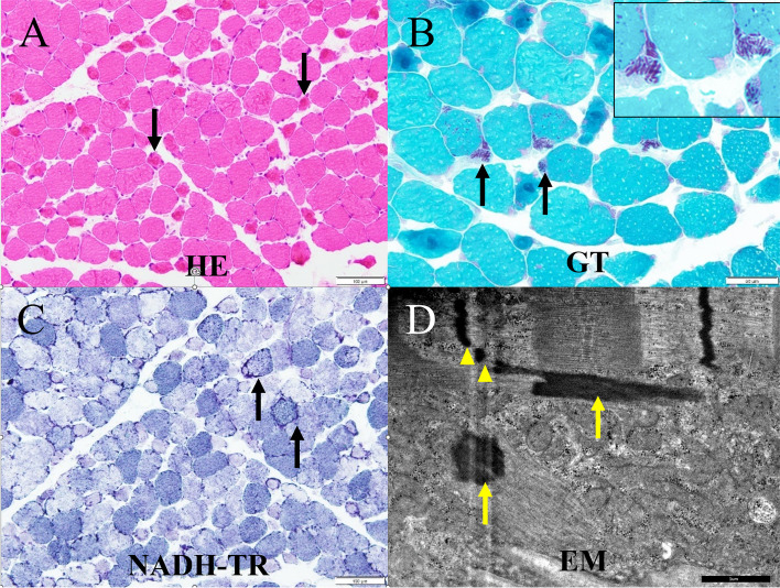
Fig. 2.
Histopathology of biopsied biceps brachii muscle. A Hematoxylin and eosin (HE) staining revealed many degenerating or atrophied muscle fibers (arrows) but no necrotic fibers. B Modified Gomori–Trichrome (GT) staining revealed nemaline rods (arrows) in some fibers and abnormal cytoplasmic agglutination (arrow heads) within atrophied muscle fibers. C Nicotinamide adenine dinucleotide tetrazolium reductase (NADH-TR) staining revealed a lobulated appearance (arrows) and focally increased or decreased oxidative enzyme activity. D Electron microscopy (EM) indicated that nemaline bodies (arrows) were of the same electron density as the Z line, with some bodies being physically connected to the Z line (arrow heads)

