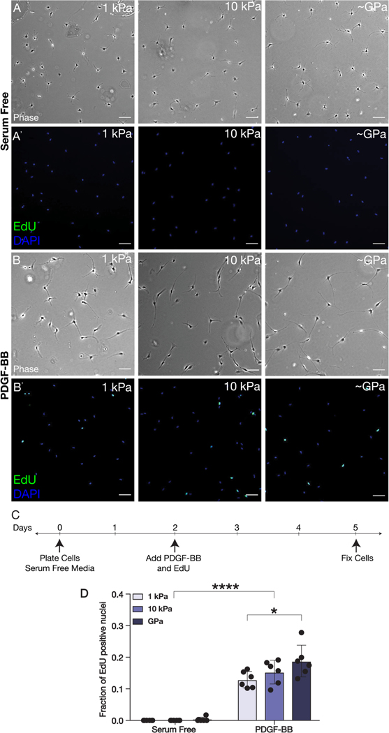
Fig. 2: PDGF-BB-mediated proliferation is modulated by substratum stiffness.
(A-B) Representative wide-field fluorescence and (A’-B’) phase contrast images of corneal keratocytes cultured on substrata of different stiffnesses (1 kPa, 10 kPa, ~GPa) and stained for EdU (green) and DAPI (blue). Cells were cultured in either (A-A’) serum-free conditions or (B-B’) in medium containing PDGF-BB. Scale bars, 100 μm. (C) Experimental timeline. Cells were pulsed with EdU and treated with PDGF-BB for 72 hr. (D) Quantification of EdU incorporation. Error bars represent mean ± s.d. for n = 6 substrates from 3 experimental replicates. A two-way ANOVA with a Tukey post-hoc test was used to evaluate significance among groups (*, p < 0.05; ****, p < 0.0001).

