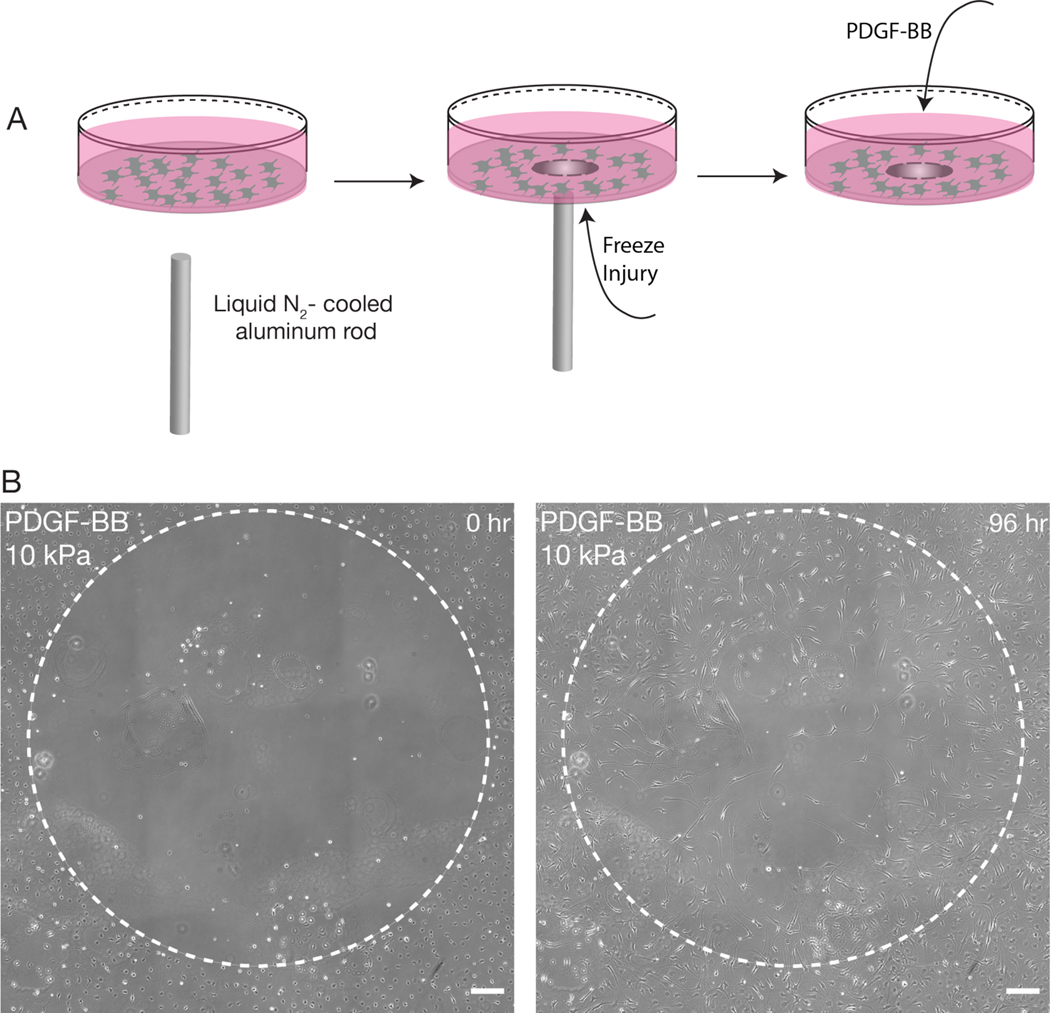
Fig, 3: Freeze injury assay induces a circular decellularized wound on PA substrata.
(A) Schematic overview of freeze injury experiment. A liquid nitrogen-cooled rod is used to create a circular freeze injury within a confluent monolayer of corneal keratocytes. (B) Representative phase contrast images of a freeze injury on a stiff (10 kPa) PA substratum after 0 and 96 hr of time-lapse culture. Note that keratocytes have migrated into the decellularized region. Scale bar, 200 μm.

