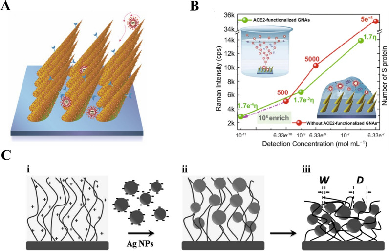
Fig. 4.
SERS biosensor based on peptide. A Schematic diagram of “virus traps” nanostructure SERS sensor for capturing SARS-CoV-2. [6] B Intensity of Raman bands (1027 cm−1) of SARS-CoV-2 S protein with different concentration detected with ACE2 functionalized GNAs and without ACE2 functionalized GNAs. The value marked on the line represents the number of S proteins in one Raman focused window. η represents enrichment multiple by ACE2. [6] C Schematic illustration of the fabrication of Ag NP-t-PLL film. (i) The amine groups of PLL chains of the t-PLL brush exposed positive charges in Ag NPs solution. The negatively charged Ag NPs were conjugated onto the film via strong electrostatic interaction and thus the (ii) Ag NP-t-PLL film in solution was formed. The film was removed from Ag NPs solution and then washed by deionized water. After the film was dried, (iii) the Ag NP-t-PLL film was prepared, and the W and D of Ag NP-t-PLL film were also defined. [90] A and B reprinted with permission from Ref. 6,
© 2021, Springer Nature. C reprinted with permission from Ref. 90, © 2009, American Chemical Society

