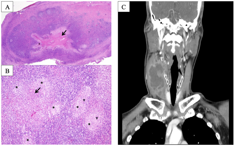
Figure 1. Photomicrographs of Case 1 and CT imaging of Case 2.
(A) H&E stain, 20x shows a section from the cervical lymph node showing effacement of normal architecture by chronic granulomatous inflammation with central necrosis (arrow). (B) H&E, 100x. High magnification showing granulomas (asterisks) consisting of epithelioid histiocytes surrounded by chronic inflammatory cells with central necrosis (arrow). Occasional multinucleated giant cells are seen (arrowheads). Ziehl-Neelsen stain does not highlight any acid-fast bacilli and the PAS stain is negative for fungal bodies. (C) CT imaging of the neck of Case 2 demonstrated a multiloculated collection in the right posterior cervical space with a hypodense center
CT: computed tomography

