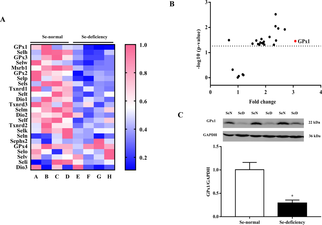
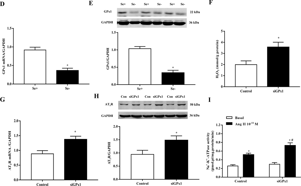
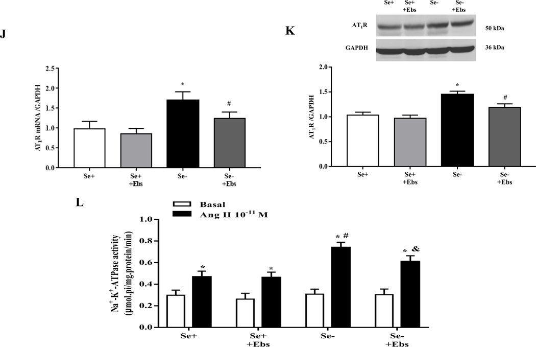
Fig. 3. Role of GPxl in the increased renal AT1R expression in selenium-deficient rats.
(A, B) Heatmap (A) and volcano plot (B) of 24 selenoproteins mRNA expression in the renal cortex of SD rats fed the selenium-deficient diet for 16 weeks. (C) The protein expression of GPx1 in the renal cortex of selenium-deficient rats (*P < 0.05 vs Se-normal, n=5/group). SeN, Se-normal; SeD, Se-deficiency. (D, E) The mRNA (D) and protein expression (E) of GPx1 in WKY RPT cells incubated with selenium-free media for 48 hours (*P < 0.05 vs Se+, n=4–5/group). Se-, selenium-deficient cells; Se+, selenium-replete cells. (F) The levels of hydrogen peroxide (H2O2) in selenium-replete RPT cells treated with GPx1 siRNA for 48 hours (*P < 0.05 vs control, n=5/group). (G, H) The mRNA (G) and protein expression (H) of AT1R in selenium-replete RPT cells treated with GPx1 siRNA (S) for 48 hours (*P < 0.05 vs control (C), n=4–5/group). (I) Effect of Ang II on Na+-K+-ATPase activity in RPT cells treated with GPx1 siRNA. RPT cells were incubated with GPx1 siRNA for 48 hours, and then treated with Ang II (10−11 M) for 30 minutes (*P < 0.05 vs basal; #P < 0.05 vs Ang II in control group, n=5/group). (J, K) The mRNA (J) and protein expression (K) of AT1R in RPT cells with selenium-free incubation and a GPX1 mimic ebselen (Ebs) treatment (30 μm) for 48 hours. AT1R mRNA and protein levels were normalized using GAPDH (*P < 0.05 vs Se+, n=4–5/group). (L) Effect of Ang II on Na+-K+-ATPase activity in selenium-deficient RPT cells treated with ebselen. Cells were incubated with selenium-free media and ebselen (30 μm) for 48 hours and then treated with Ang II (10−11 M) for 30 minutes (*P < 0.05 vs basal, n=5; #P < 0.05 vs Ang II in Se+ group, n=5/group).


