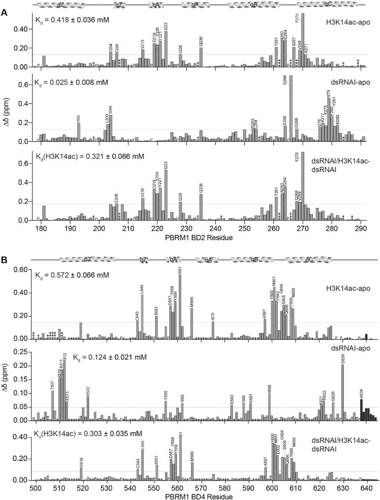
Figure 4.
BD2 and BD4 have partially overlapping nucleic acid and histone binding pockets. (A, B) Normalized chemical shift changes (Δδ) between apo and H3K14ac-bound (top), apo and dsRNAI-bound (middle), and dsRNAI-bound and H3K14ac-bound (bottom) for BD2 (A) and BD4 (B). Protein:dsRNAI, protein:H3K14ac, protein:dsRNAI:H3K14ac are in molar ratio of 1:6, 1:10 and 1:6:10 respectively for BD2. Protein:dsRNAI, protein:H3K14ac, protein:dsRNAI:H3K14ac are in molar ratio of 1:10, 1:40 and 1:10:15 respectively for BD4. Residues that are unassigned, merged with the addition of ligand, or broaden beyond detection upon addition of ligand, are marked as (*), (**) and (***), respectively. The secondary structure of the BD2 is denoted above the plots, and residues that were perturbed greater than the average plus two standard deviations (grey line) after trimming off the top 10% of CSPs are labelled.

