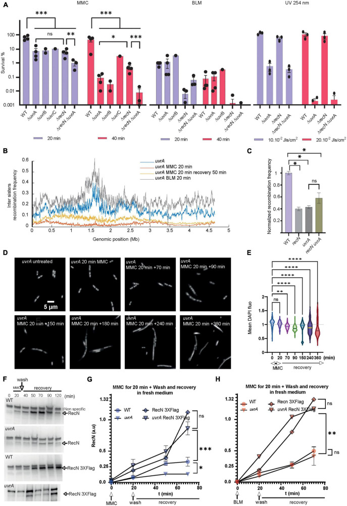
FIGURE 5.
Role of the NER in the GSR associated with BLM and MMC lesions. (A) CFU analysis of the WT, recN, uvrA, uvrB, uvrC single, and double mutants in the presence of MMC, BLM, and UV irradiation. Anova statistical test *<0.05, **<0.005, ***<0.0005, ****<0.00005. (B) Hi-SC2 profile of sister chromatid interactions in the uvrA mutants in the presence or absence of MMC and BLM. (C) Measure of inter sister locus recombination, laclox assay at the aidB locus (Vickridge et al., 2017), in the WT, recN, uvrA, and recN uvrA mutants. (D) Nucleoid imaging of the uvrA mutant during the recovery period of MMC injury. (E) Quantification of the mean DAPI fluorescence in the nucleoid area of uvrA mutant treated with MMC. Anova statistical test *<0.05, **<0.005, ***<0.0005, ****<0.00005. (F) Western blot showing RecN and RecN 3XFlag amount in WT and uvrA cells during GSR recovery. (G) Quantification of RecN amount in 3 western blot experiments performed as in panel F in the WT and uvrA mutant during the recovery period of MMC injury. RecN amount was normalized to the amount of RNA polymerase β in each sample. (H) Measure of RecN and RecN 3X-flag turnover in the WT and uvrA mutant during the recovery period of BLM injury (N = 3 western blots performed in identical conditions).

