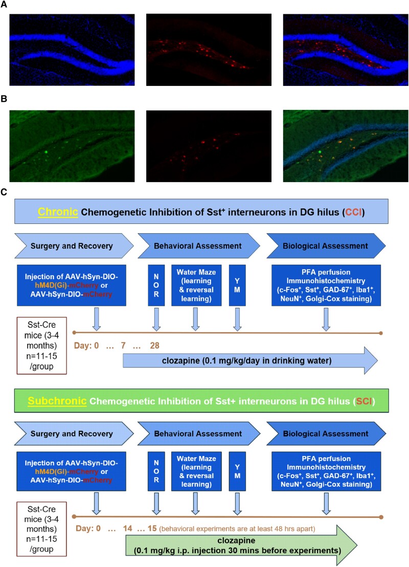
Fig. 1.
Experimental design of DG hilus Sst+ cell manipulation. A) Immunofluorescence staining of coronal sections from an AAV-hSyn-DIO-mCherry mouse showing DAPI counterstain (left: DAPI; middle: mCherry expression; right: merged image). B) Immunofluorescence colabeling of coronal sections from an AAV-hSyn-DIO-mCherry mouse (left: Sst labeling with Alexa Fluor 488; middle: mCherry; right: merged image, with DAPI). C) Top: chronic chemogenetic inhibition of DG hilus Sst+ cells. Bottom: subchronic chemogenetic inhibition of DG hilus Sst+ cells.

