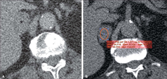
Figure 2.

A 53-year-old female patient. A: Axial contrast-enhanced CT scan, in the venous phase, showing a right-sided homogeneous adrenal lesion measuring 2.8 cm (asterisk). B: Axial unenhanced CT scan at the same level with an ROI drawn at the center of the lesion, showing a mean attenuation of 24.7 HU, which is suggestive of an LPA (although not meeting the criteria at this point). However, the histogram analysis P10 and calcP10 showed that there was more than 10% negative voxels, further suggesting a diagnosis of LPA. At this writing, the patient is asymptomatic and the lesion has been stable for 62 months.
