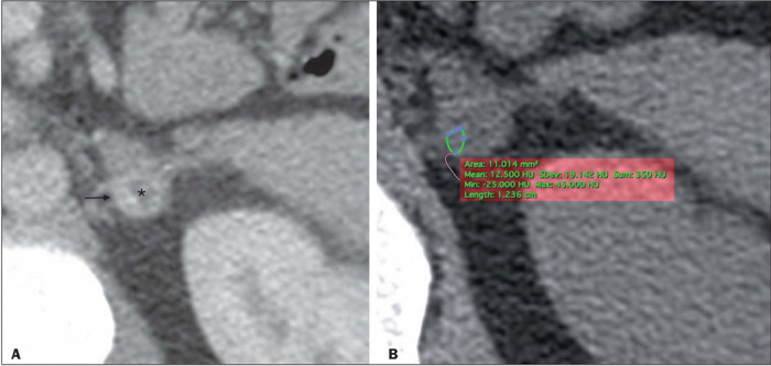
Figure 4.

A 40-year-old male patient with symptoms and laboratory test results indicative of a PCC. A: Contrast-enhanced CT scan, in the arterial phase, showing a hypervascular 1.5-cm lesion on the left adrenal gland (arrow) with a central cystic area (asterisk). B: Axial unenhanced CT scan at the same level with an ROI drawn to avoid the cystic portion. The mean attenuation was 12.5 HU. The histogram analysis P10 and calcP10 showed more than 10% negative voxels. Aside from the clinical and biochemical setting, this could be assumed to be an AA, based on histogram analysis and calcP10 criteria, were it not for the heterogeneity of the lesion.
