Figure 4:

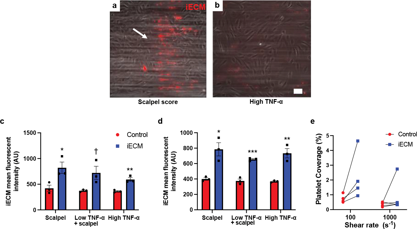
iECM binds to injured endothelial cells and facilitates platelet adhesion in vitro. Representative images of iECM binding to either scalpel score (white arrow region) damaged HUVECs (a) or strongly TNF-α inflamed HUVECs (b) under flow conditions with red blood cells and plasma in an in vitro adhesion flow assay Scale bar is 100 μm. iECM binding to injured (scalpel) and/or inflamed (TNF-α) HUVECs was significantly increased over controls at both low (100s−1) (c) and high (1000s−1) (d) shear rates (n=3 all groups). Low shear (c): scalpel: *p=0.03, low TNF-α + scalpel: †p=0.055, high TNF-α: **p=0.004. High shear (d): scalpel: *p=0.01, low TNF-α + scalpel: ***p<0.001, high TNF-α: **p=0.004. Under flow conditions with whole blood, iECM increased platelet adhesion for all 4 blood donors at low shear rates (e). Data are mean ± SEM. All data are biological replicates. Data were evaluated with a two tailed unpaired t-test.
