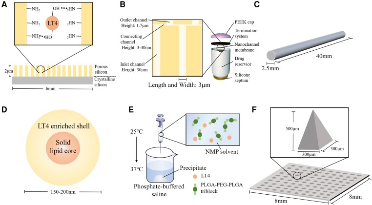
Figure 7.
(A) The structure of 3-(aminopropyl)triethoxysilane-functionalized, oxidized porous silicon film. The LT4 molecules bind to porous silicon with hydrogen bonding. (B) The rendering of nanofluidic drug delivery implants and the cross section of microchannels on nanofluidic membranes. The implant is ∼2.5 cm in length. The silicon nanochannel membranes are 6 × 6 mm wide and 730 µm in height. The length and width of the 3 channels (inlet, connecting and outlet) are 3 µm. (C) The rendering of poly(caprolactone)-based subcutaneous implant. (D) The rendering of LT4-loaded poly ethylene glycol 100 stearate–coated solid lipid nanoparticles. (E) The process of sol to gel transformation. Thermosensitive triblock undergoes polymerization. (F) The rendering of microneedle patch. Abbreviation: PEEK, polyether ether ketone. The figure was partly generated using illustrative elements from Servier Medical Art, provided by Servier, licensed under a Creative Commons Attribution 3.0 unported license.

