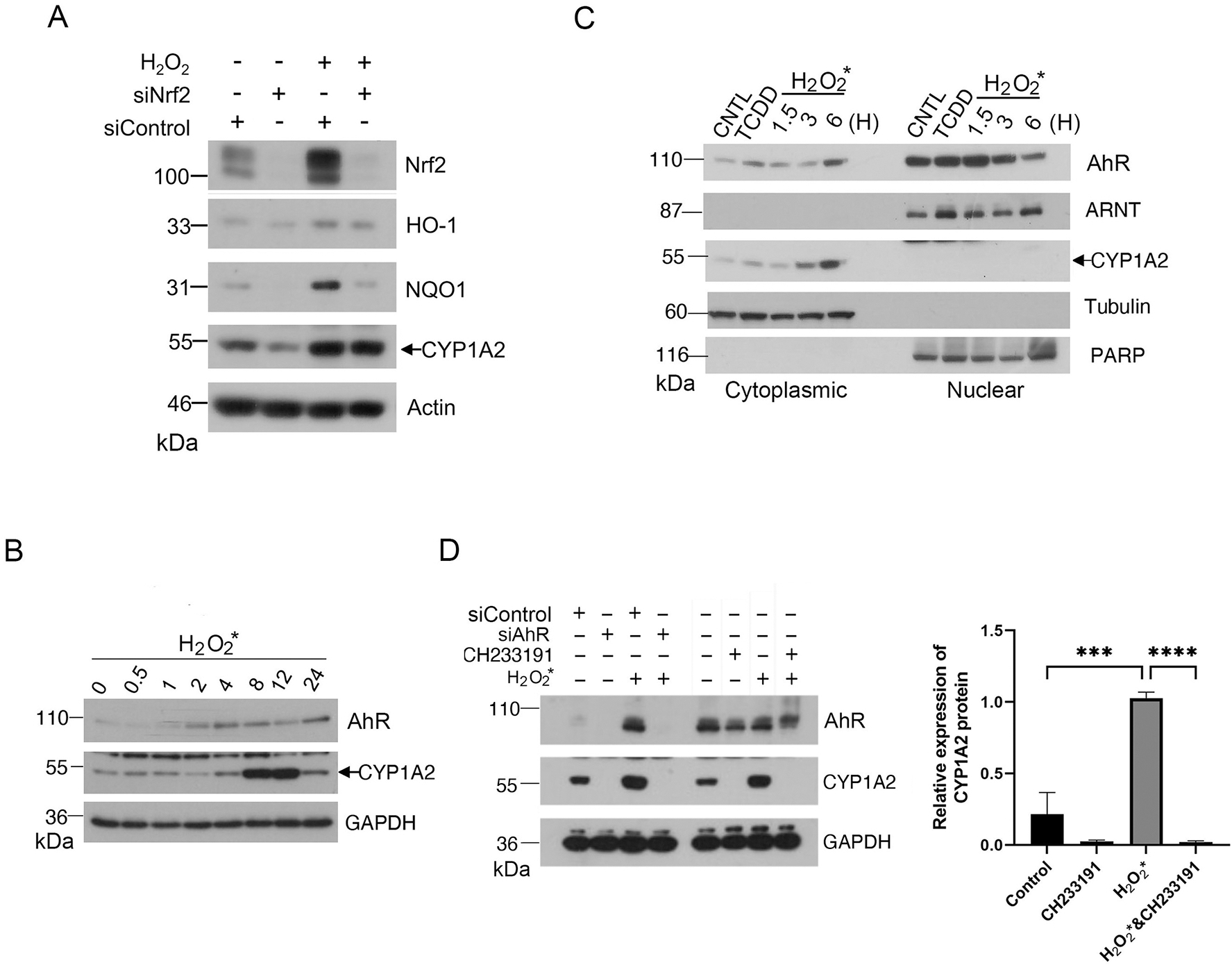
Figure 3.

Oxidant H2O2 induces an AhR agonist(s) in the culture medium. (A) HaCaT cells were transfected with Nrf2-specific siRNAs or control siRNAs for 24 h before treatment with H2O2 for 24 h. At the end of experiments, cells were lysed, and equal amounts of cell lysates were blotted for Nrf2, HO-1, NQO1, CYP1A2, and actin. (B) H2O2 (at a final concentration of 0. 2 mM) was supplemented to the warmed culture medium (DMEM with FBS) for 30 min in dark before it (H2O2-containing medium) was added to HaCaT cells. Cells were incubated with H2O2-containing medium for various times before they were harvested for lysis. Equal amounts of cell lysates were blotted for AhR, CYP1A2 and GAPDH. (C) HaCaT cells were treated with H2O2 for 1.5, 3, or 6 h before they were collected for fractionation into the cytoplasmic or nuclear parts. Cells treated with TCDD (5nM) or vehicle were also fractionated as control. Equal amounts of cell lysates of various treatments were blotted for AhR, ARNT, CYP1A2. Cell lysates were also blotted for compartmental markers including α-tubulin (cytoplasmic) and PARP-1 (nuclear). (D) HaCaT cells were transfected with AhR siRNAs or control siRNAs for 48 h before they were cultured in H2O2-pretreated medium for 8 h. HaCaT cells incubated in H2O2-pretreated medium were also treated with CH233191 or vehicle for 8 h. At the end of cultures, cells were collected and lysed. Equal amounts of cell lysates were blotted for AhR, CYP1A2 and GAPDH. Western blot (left) is representative of one experiment and triplicated western blot quantifications (right) were analyzed by statistical analysis as described Methods.
