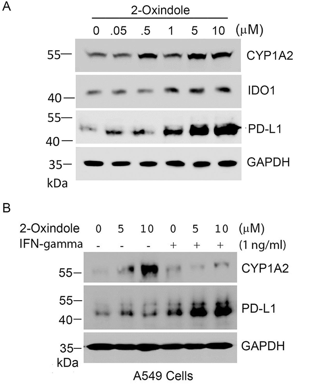
Figure 6.

Oxindole induces expression of PD-L1. (A) A549 cells were treated with 2-oxindole of various concentrations as indicated for 24 h after which cells were lysed. Equal amounts of cell lysates were blotted for CYP1A2, IDO1, PD-L1, and GAPDH. (B) A549 cells were treated with 2-oxindole (5 and 10 μM) and/or IFN-γ (1 ng/ml) as indicated for 24 h. Equal amounts of cell lysates were blotted for CYP1A2, PD-L1, and GAPDH.
