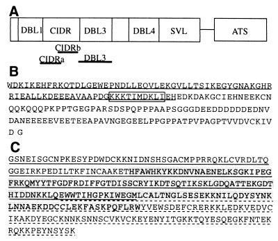
FIG. 1.
(A) Schematic representation of the location of the CIDR and DBL3 domains of the CS2 var gene. (B) Amino acid sequence of the CIDRb protein, showing a putative CSA binding motif (boxed) and an overlap with a region associated with CD36 binding in other isolates (underlined). (C) Amino acid sequence of the DBL3 protein and the smaller DBL3-5′ (underlined), DBL3-C (highlighted), and DBL-3′ (dashed underline) polypeptides.

