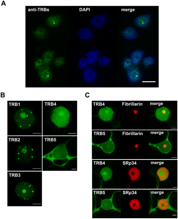
Fig. 5.
Subcellular localization of native TRBs and GFP-TRB fusion proteins. (a) Isolated nuclei from A. thaliana seedlings were subjected to immunofluorescence using an anti-TRB antibody combined with DAPI staining. All five native members of the TRB family are visualized. Scale bar = 10 µm. (b) TRB1-5 were fused with GFP (N-terminal fusions), expressed in N. benthamiana leaf epidermal cells and observed by confocal microscopy. Single images of areas with nuclei are presented. Scale bars = 5 µm. (c) Co-localization of TRB4 and TRB5 (N-terminal GFP fusions) with a nucleolar marker (Fibrillarin-mRFP) and a nucleoplasm marker (SRp34-mRFP) was performed as described in B). Single images of areas with nuclei are presented. Scale bars = 5 µm

