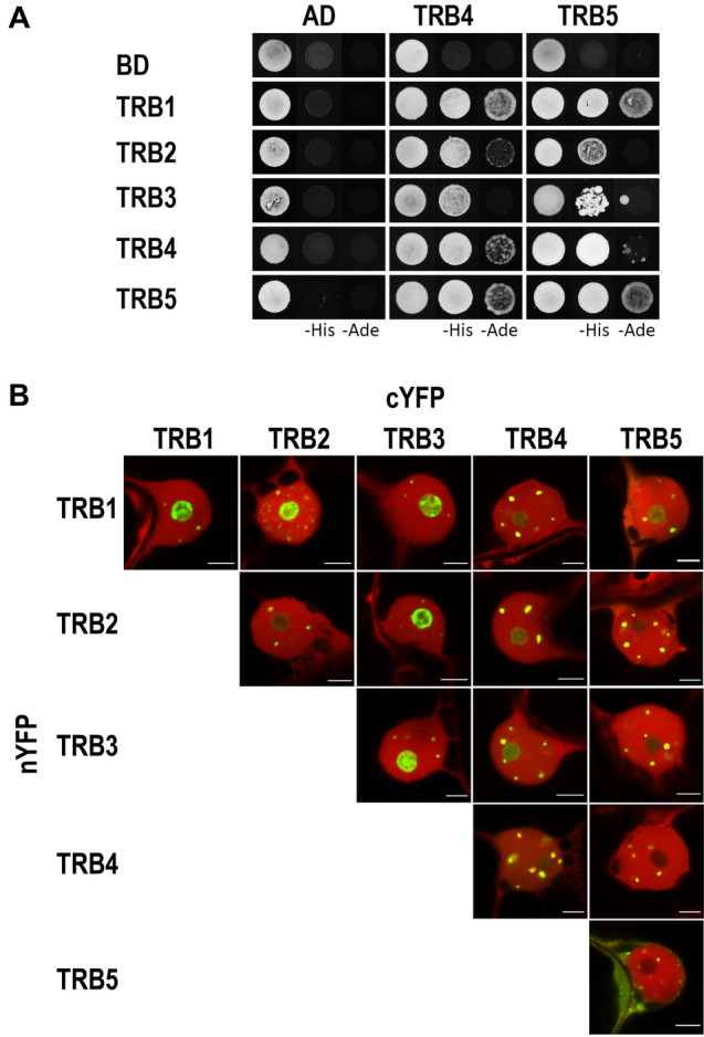
Fig. 6.

Dimerization of TRB proteins. (a) The Y2H system was used to assess mutual protein–protein interactions of TRBs. Two sets of plasmids carrying the indicated protein fused to either the GAL4 DNA-binding domain (BD) or the GAL4 activation domain (AD) were constructed and introduced into yeast strain PJ69-4a carrying reporter genes His3 and Ade2. Interactions were detected on histidine-deficient SD medium (–His), or under stringent adenine-deficient SD medium (–Ade) selection. Co-transformation with an empty vector (AD, BD) served as a negative control. (b) Interactions of TRBs fused with nYFP or cYFP part were detected using the Bimolecular fluorescence complementation (BiFC) assay in N. benthamiana leaf epidermal cells. Shown here are single images of merged signals of reconstructed YFP (interaction of the tested proteins) and signals of mRFP (internal marker for transformation and expression) fluorescence detected by confocal microscopy. For separated fluorescent emissions, see Supplemental Fig. 5. Scale bars = 5 µm
