Figure 1.
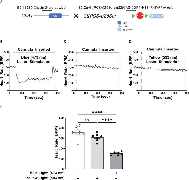
Selective activation of DMN cholinergic neurons invokes efferent vagus nerve activity, reducing heart rate. (A) Breeding strategy used to generate ChAT-ChR2-YFP mice which express a photosensitive channelrhodopisin in ChAT-positive cells, including cholinergic neurons. (B–D) Optogenetic stimulation of DMN cholinergic neurons produced a significant decrease in heart rate in ChAT-ChR2-eYP mice. A fiber optic cannula was placed in the left DMN of ChAT-ChR2-YFP mice and animals were then subjected to (B) blue light (473nm, 20Hz, 25% duty cycle, 8-12 mW, 5 minutes), (C) no light, or (D) yellow light (593.5nm, 20Hz, 25% duty cycle, 8-12 mW, 5 minutes) stimulation. A decrease in heart rate was observed during blue laser stimulation. Representative heart rate data is shown for each condition. (E) Stimulation of ChR2-expressing DMN cholinergic neurons with blue light, but not with yellow light, induces bradycardia in ChAT-ChR2-eYFP mice. Data are represented as individual mouse data points, which represent an average heart rate over the stimulation period, with mean ± SEM. One-way ANOVA with Tukey’s multiple comparison test, ****P ≤ 0.0001, ns, not significant.

