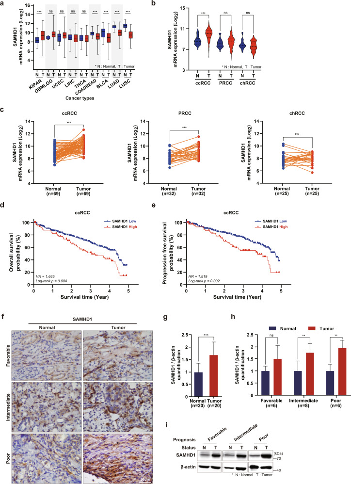
Fig. 1. High SAMHD1 expression is associated with poor prognosis in ccRCC patients.
a Comparing the gene expression of SAMHD1 in various cancer types using the TCGA database. KIPAN, pankidney cancer; GBMLGG, glioma; UCEC, uterine corpus endometrial carcinoma; LIHC, liver hepatocellular carcinoma; THCA, thyroid carcinoma; COADREAD, colorectal adenocarcinoma; BLCA, bladder urothelial carcinoma; LUAD, lung adenocarcinoma; LUSC, lung squamous cell carcinoma. b Comparison of SAMHD1 mRNA expression between tumor and normal tissues according to histological RCC subtypes. ***p < 0.001. c Comparative mRNA expression of SAMHD1 between paired tumor and nontumor tissues according to histological RCC subtypes. ***p < 0.001. d Kaplan–Meier analysis of 5-year overall survival (OS) between SAMHD1 high expression (n = 77) and SAMHD1 low expression groups (n = 303). Log-rank p = 0.004. e Kaplan–Meier analysis of progression-free survival (PFS) between SAMHD1 high expression (n = 78) and SAMHD1 low expression groups (n = 300). Log-rank p = 0.002. f Immunohistochemistry (IHC) of SAMHD1 in ccRCC tissues. Scale bar, 50 μm. g Immunoblotting was used to analyze the protein level of SAMHD1 in 20 paired cancer and normal tissues. SAMHD1 protein levels were normalized to corresponding β-actin levels. **p < 0.01. h SAMHD1 protein levels normalized to β-actin levels in ccRCC tissues according to patient prognosis were analyzed by western blotting. *p < 0.05. Data are presented as the mean ± SD. i Representative western blot bands are presented with the corresponding prognosis.

