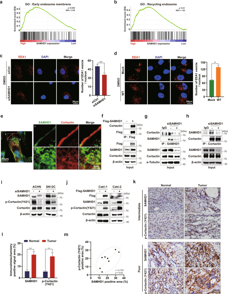
Fig. 6. SAMHD1 participates in endosome formation and binds to cortactin directly.
a, b GSEA analysis of SAMHD1 mRNA levels and endocytosis-related signaling pathways. c ACHN cells were transfected with control or siSAMHD1 and immunolabeled for EEA1. The nuclei were stained with DAPI. Scale bar, 20 μm. Graphs present the number of EEA1-expressing vesicles per single cell (n = 55 cells). Data are presented as the mean ± SEM. The experiments were independently performed three times. ***p < 0.001. d SAMHD1-overexpressing Caki-1 cells were immunolabeled for EEA1. The number of EEA1-expressing vesicles per single cell is shown (n = 55 cells). Scale bar, 20 μm. Data are presented as the mean ± SEM. The data shown are representative of three independent experiments. ***p < 0.001. e SAMHD1 colocalized with cortactin in the cytoplasmic circular-shaped organelles and on the lamellipodia. f Exogenous Flag-tagged SAMHD1 was immunoprecipitated with magnetic Flag beads from Flag-SAMHD1-overexpressing Caki-1 cells. g Endogenous SAMHD1 was immunoprecipitated from SAMHD1-knockdown SN12C cells. Anti-cortactin antibodies were used to examine SAMHD1 and cortactin binding. IgGs were incubated with cell lysates as negative controls. h Endogenous cortactin was immunoprecipitated from SAMHD1-downregulated ACHN cells. Anti-SAMHD1 antibody was used to examine SAMHD1 and cortactin binding. IgG was incubated with cell lysates as a negative control. i, j Phosphorylated cortactin (Y421) and total cortactin levels were evaluated using western blotting in SAMHD1-knockdown ACHN and SN12C cells or SAMHD1-overexpressed Caki-1 and Caki-2 cells. k IHC analysis of SAMHD1 and phosphorylated cortactin (Y421) in ccRCC patient tissues. Scale bar, 50 μm. l Areas with positive IHC staining were measured and are shown on the graph. Data are presented as the mean ± SD. ***p < 0.001; **p < 0.01. m Correlation between SAMHD1 and phosphorylated cortactin (Y421) expression from IHC analysis of ccRCC patient tissues.

