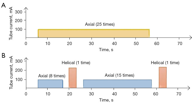
Figure 1.
The scheme of the 2 CT scanning methods in the study. (A) The continuous perfusion scanning pattern. (B) The intermittent perfusion scanning pattern. The continuous perfusion scan method used axial patterns, and the Z-axis scanning length was about 150 mm. The tube voltage was 100 kV, and the tube current was 100 mA. Each scan time was 2 seconds, and the total scanning time was 50 s (25 passes). The intermittent perfusion scan method started with helical scanning after 20.5 s (perfusion scanning occurred 8 times after 6 s during the injection of the nonionic contrast agent). The time of the helical scanning and conversion between the helical mode and axial mode was about 8.6 to 13.7 s. The helical scanning was arterial phase scanning. Then, perfusion scanning occurred 13 times for the whole pancreas in the axial pattern, which was then converted to the helical pattern for venous phase scanning. CT, computed tomography.

