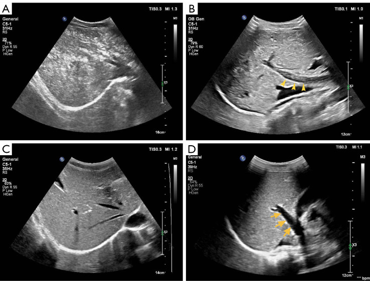
Figure 3.
A 42-year-old male patient was intravenously injected with 3.0 mL of SonoVue® to discriminate malignant thyroid nodules. The patient presented with mild abdominal discomfort after 32 minutes of CEUS. (A) At the same time, scattered tiny hyperechoic spots were detected in the liver. (B) Flowing hyperechoic spots were observed in the portal vein (arrows) (C) After 2 hours of SonoVue® injection, the patient’s abdominal discomfort was relieved, and the hyperechoic areas in the liver almost disappeared. (D) No hyperechoic spots were found in the portal vein (arrows). CEUS, contrast-enhanced ultrasound.

