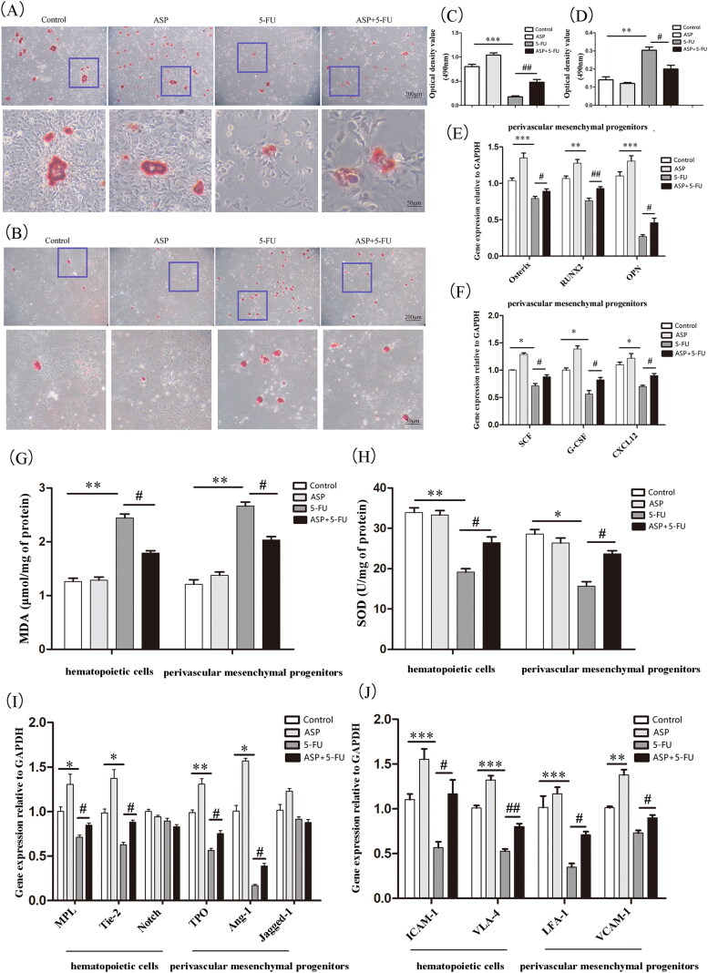
Figure 3.
Angelica sinensis polysaccharides protected the hematopoietic niche function of perivascular mesenchymal progenitors. (A) The representative image of induced osteogenic differentiation of perivascular mesenchymal progenitors in vitro is visualized by alizarin red staining. (B) The representative image of induced adipogenic differentiation in vitro of perivascular mesenchymal progenitors is stained by oil red O. (C) Alizarin red dye was dissolved in isopropyl alcohol, and the absorbance was measured at 490 nm, and quantitative analysis was performed by Image J software (n = 3). (D) The absorbance of dissolved oil red O dye at 490 nm was analyzed by Image J software. (n = 3) (E) Quantitative analysis of osteogenic-related mRNA expression (n = 3). (F) Quantitative analysis of hematopoiesis growth factor mRNA expression (n = 3). (G) Analysis of malondialdehyde (MDA) levels in perivascular mesenchymal progenitors and co-cultured hematopoietic cells. (H) Analysis of superoxide dismutase (SOD) levels in perivascular mesenchymal progenitors and co-cultured hematopoietic cells. (I) Quantitative analysis of mRNA relative expression of hematopoietic cell and perivascular mesenchymal progenitors interaction signaling molecules (n = 3). (J) Analysis of mRNA expression of adhesion molecules between hematopoietic cells and perivascular mesenchymal progenitors. (***p < 0.001 **p < 0.01 *p < 0.05 vs. control group; ##p < 0.01 #p < 0.05 vs. 5-fluorouracil (5-FU) group, n = 3).

