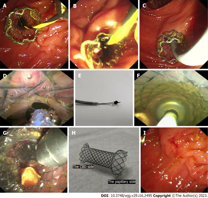
Figure 1.
The procedures of cholangioscopy-assisted extraction through novel papillary support for small-calibre and sediment-like common bile duct stones. A: The novel papillary support was placed in the lower common bile duct (CBD) and papilla; B: Many sediment-like CBD stones flowed from the support under endoscopic aspiration; C: The cholangioscope (Micro-Tech, eyeMax) was inserted into the CBD; D: The single clumpy CBD stone was collected by the basket under cholangioscopy; E: The stone, collected by the basket, was extracted from the CBD and body along with the cholangioscope; F: Multiple clumpy CBD stones were held by the balloon under cholangioscopy; G: One stone, held by the balloon, was extracted from the CBD along with the cholangioscope; H: The novel single dumbbell-style papillary support was removed from the body; I: Papillary morphology immediately after the removal of papillary support.

